Diagnostic Imaging
X-rays
At Zoo Veterinary Center, we provide advanced diagnostic imaging services tailored to ensure the comprehensive care of your beloved pets. Our state-of-the-art X-ray and ultrasound technologies enable our veterinarians to accurately diagnose and treat a wide range of medical conditions.
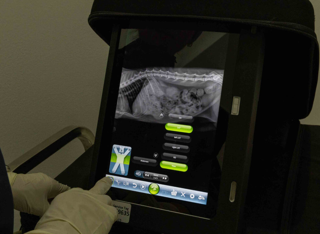
X-ray (Radiography): X-ray imaging is a vital tool in veterinary medicine, allowing us to visualize the internal structures of our pets. It helps in diagnosing fractures, joint abnormalities, foreign bodies, and certain organ conditions. Our digital X-ray equipment produces high-quality images swiftly, ensuring minimal stress for your pet.
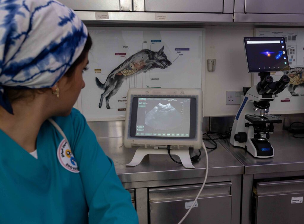
Ultrasound (Ultrasonography):
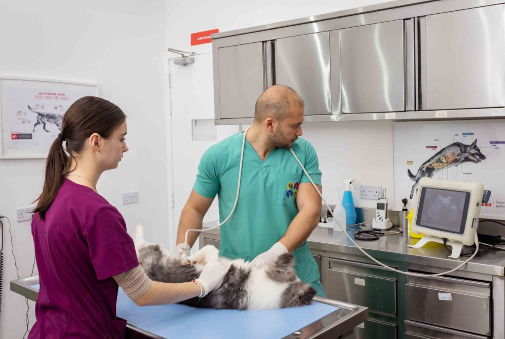
Ultrasound technology utilizes sound waves to create real-time images of your pet’s internal organs. This non-invasive technique is particularly useful for assessing soft tissues and organs such as the heart, liver, kidneys, and reproductive organs. It aids in diagnosing conditions like tumors, cysts, and abnormalities in blood flow.
Why Choose Us for Diagnostic Imaging?
- Expertise: Our veterinarians are experienced in interpreting diagnostic images to provide accurate diagnoses and treatment plans.
- Compassionate Care: We prioritize your pet’s comfort and safety throughout the imaging process.
- Comprehensive Approach: Combined with our other diagnostic and treatment services, X-ray and ultrasound help us deliver thorough care tailored to your pet’s needs.
Whether your pet needs routine screening or specialized diagnostic imaging, Zoo Veterinary Center is here to provide exceptional veterinary care. Contact us today to schedule an appointment or to learn more about our services.
Commonly Asked Questions
Diagnostic imaging refers to techniques like X-rays and ultrasound that create visual images of the inside of your pet’s body. These images help veterinarians diagnose and evaluate a wide range of conditions, from fractures and tumors to internal injuries and organ abnormalities. X-rays are particularly useful for viewing bone structures and detecting issues like broken bones or arthritis, while ultrasound provides detailed images of soft tissues and organs, helping to identify problems with the heart, liver, kidneys, and more. These imaging techniques are essential for accurate diagnosis and effective treatment planning.
Yes, both X-rays and ultrasounds are generally safe for pets. X-rays use a small amount of radiation to create images, but the levels are minimal, and the risk is low. Veterinary clinics take precautions to minimize radiation exposure, such as using protective gear and only performing X-rays when necessary. Ultrasound, on the other hand, uses sound waves rather than radiation, making it completely safe and non-invasive. Both imaging methods provide valuable diagnostic information with minimal risk to your pet.
For an X-ray, your pet will typically need to be positioned in a specific way to obtain clear images. The procedure is quick, but your pet may need to be held still, which sometimes requires mild sedation, especially for anxious or uncooperative pets. For an ultrasound, your pet may need to be sedated or kept still to get clear images, though this is less common. The procedure involves applying a special gel to the skin and using a probe to capture images. Both procedures are generally brief, and your veterinarian will discuss the results with you once the images are reviewed.